

The selection, incision, and suturing of the donor area during a hair transplant is an issue that concerns candidates for hair implantation with the FUT technique. Read everything you need to know about trichophytic closure..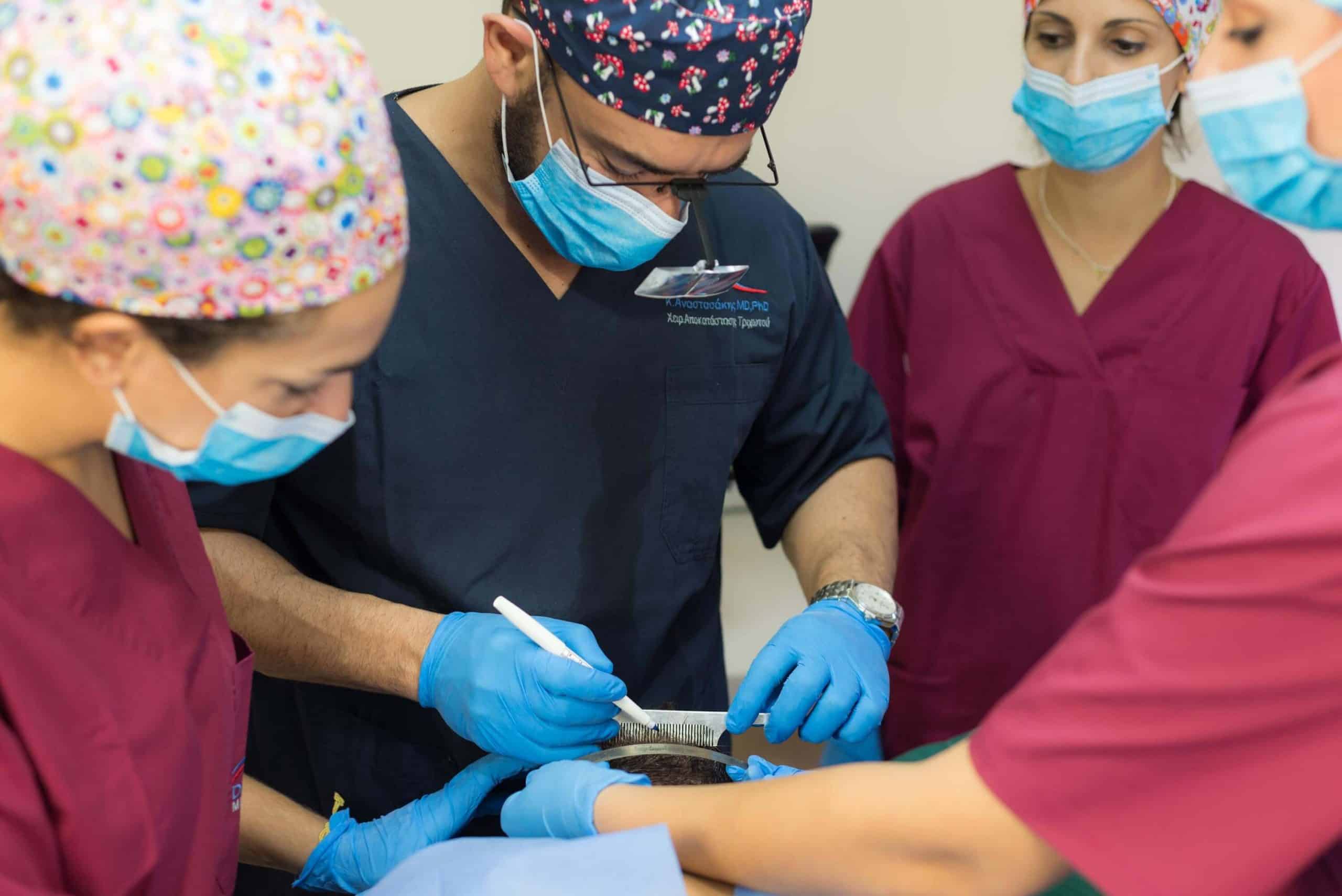
Donor Area Suturing: Key Concepts
- During the removal of the strip graft from the donor area, the goal is to minimize tissue damage and follicular loss. Careful surgical technique, along with innovative atraumatic methods, is essential.
- Closure of the donor area defect can be performed either with sutures or staples, depending on the surgeon’s preference and the specifics of each case.
- With proper surgical technique and attentive postoperative care by the patient, the scar width typically ranges from 0.3–0.7 mm, and maximum attention must be given to minimizing follicular loss on both sides of the incision.
- Trichophytic closure is an advanced suturing technique of the donor area, in which, during healing, hairs grow through the scar, making it virtually undetectable.
The process of removing a strip of scalp from the occipital (donor) area inevitably causes partial or complete transection of a number of hair follicles along the edges of the incision. This trauma results either in their necrosis in situ or, at best, in poor growth when transplanted into the recipient area.
Over the years, it has been proven that the rate of iatrogenic follicular loss, as well as damage to important vascular and nerve branches, can be minimized with the proper surgical technique. Let’s take a closer look at the details of these techniques:
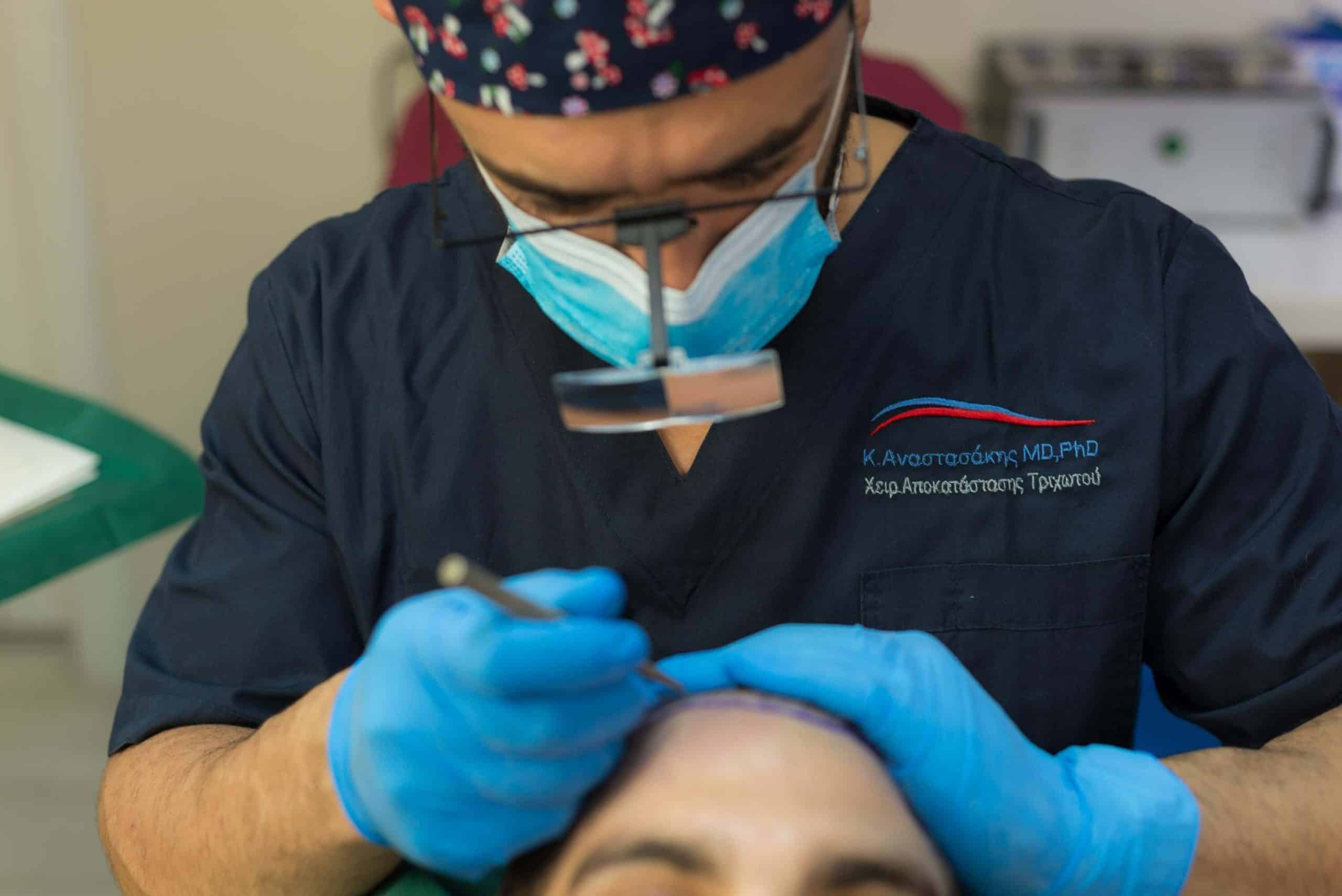
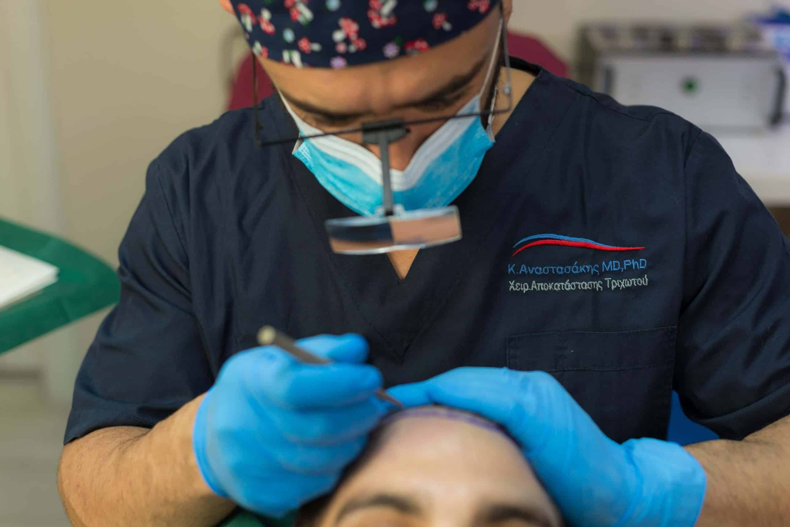
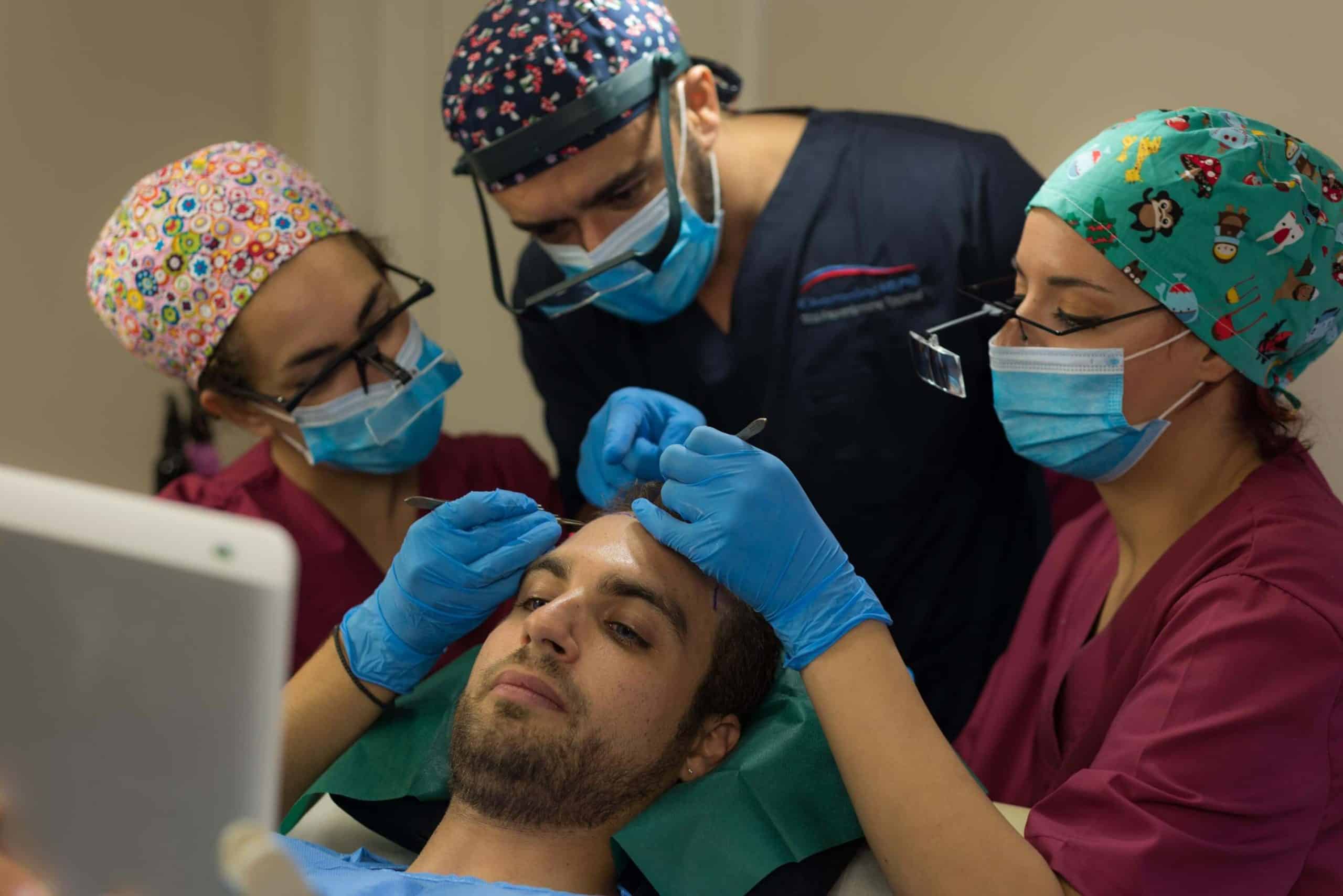
- Angle of incision: Hairs emerge from the scalp at an acute angle, which moreover differs from one area of the scalp to another, even along the occipital surface of the same individual. To reduce follicular loss, the surgical scalpel must enter in absolute parallelism with the hairs, while the surgeon must constantly check the direction of the incision and ensure that the scalpel remains parallel to the hairs.
- Tumescent anesthesia: The injection of a large quantity of diluted anesthetic and vasoconstrictor solution causes subcutaneous tension and iatrogenic edema. The intraoperative induction of temporary iatrogenic edema in the donor area results in temporary separation and elevation (verticalization) of the hairs in order to avoid injury to adjacent follicles, as well as hydraulic vasoconstriction and reduction of bleeding.
- Type of scalpel: As already analyzed in a previous section, the use of a single-bladed knife reduces the unintentional transection of follicles at the edges of the incision to 1–2%, whereas with a multi-bladed knife with three blades it is approximately 15%.
- Depth of incision and other instruments: The depth of the incision must be minimal. Specifically, recent publications describe techniques involving only scoring of the epidermis at the outline of the strip graft at a depth of just 1–2 mm. Subsequently, tissue is separated with special atraumatic instruments until it detaches from the occipital region and is removed.
Sandoval was the first to describe such a technique in 2005. Later, Haber developed the Haber Spreader, Tykocinski initially used Mosquito forceps and later the Intruder instrument, while techniques have also been reported using Kleinert-Kuts hooks, 5″ in length and 7 mm in diameter.
With a combination of the above techniques, the injury to adjacent hair follicles can literally be reduced to zero
Donor area closure during hair transplantation
The closure of the skin defect resulting from the removal of the strip graft in FUT hair transplantation is performed either with sutures or with the use of staples. Before suturing, the bleeding vessels are usually cauterized, most commonly the “Arsenault vessel,” located in the mid-occipital region and named after the surgeon from Unger’s team who observed the high frequency with which this vessel was transected during the elliptical incision. It is important to remember that large vessels are accompanied by sensory nerves. Therefore, cauterization must be carried out with caution.
Sutures or staples in the donor area?
Many surgeons prefer wound closure with staples because of the speed of the procedure and the fact that this technique is less traumatic for the surrounding hair follicles. The use of staples provides similar effectiveness in minimizing scarring compared to non-absorbable monofilament sutures, and it is also safer for the staff since it eliminates the risk of needle-stick injuries. Contrary to common belief, staples ultimately prove to be more cost-effective when all parameters are taken into account.
However, patients often complain that staples are uncomfortable and painful during the postoperative period as well as during their removal. Unlike simple sutures, staples do not allow the surgeon to accurately assess the tension of the wound edges during closure. Nevertheless, since staples are completely inert, they can remain in the scalp for up to 15–20 days. This is an extremely useful property in cases with increased tension at the wound edges, which require sutures to remain in place for a longer period.
For suture closure, the material most commonly used is poliglecaprone 25, #5-0 or #6-0, applied in a continuous stitch with close and shallow passes. This approach minimizes the loss of hair follicles along the path of the needle and suture while distributing wound tension evenly. The use of this specific suture type has been shown to produce the lowest tissue reaction compared to other materials, such as vicryl or chromic sutures.
Other surgeons prefer to use absorbable sutures in patients who live far from the surgical site and cannot return for suture removal, as the newer absorbable monofilaments demonstrate excellent performance.
There is ongoing debate about whether it is necessary to suture the deeper tissue layers in order to support the superficial sutures and reduce surface tension that could lead to a wider scar. A two-layer closure is recommended when there is tension at the wound edges, when a strip graft wider than 1 cm is removed, and in cases where the donor area has been previously operated on.
The recommended method involves initial gentle undermining of the scalp for less than 10 mm on both edges, followed by suturing of the dermis on one side and the fascia on the other with an absorbable Poliglactin 910 suture, and finally, closure of the wound. With proper surgical technique and preoperative preparation, the width of the remaining scar measures 0.3–0.7 mm, and the scar is virtually invisible, unless one knows exactly where to look or if the patient shaves their hair very short (<5 mm).
Each surgeon uses a different suturing method for donor area closure, and the final choice depends on the specific case and factors such as scalp laxity, the width of the excised strip, and the presence of previous scars from strip excision or older scalp reduction surgeries.
In general, there is no single “ideal” closure method, and when the surgeon is well-versed in the surgical anatomy of the scalp and avoids surgical overextensions, the likelihood of complications in the donor area is significantly reduced. Nevertheless, every surgeon will encounter during their career at least one very challenging case with minimal scalp mobility—either due to previous surgeries that have altered its elasticity, or because of individual anatomical factors—a combination that can intimidate even the most experienced surgeon.
Collateral loss during donor area closure
Regardless of the closure technique and the surgeon’s skill, some follicular units (FUs) are inevitably damaged each time the donor area is sutured. One reason is that, contrary to popular belief, the intraoperative tension on the wound is not the final tension—it is only the beginning of tension.
Specifically, wounds closed under minimal tension can develop significant edema during the first postoperative days. This swelling can exert high pressure on the sutures and surrounding tissue, and in some cases, may reduce scalp perfusion, potentially leading to a wide scar and loss of follicular units along the edges of the incision.
To minimize these losses, the following basic guidelines are recommended:
-
Sutures should be very superficial and placed close to the edges of the incision.
-
Avoid continuous sutures or subdermal mattress closure, as these can cause strangulation of the follicular units.
-
When a previous scar exists, use it as the upper boundary of the incision, limiting tissue damage to one side only. Do not completely excise the old scar—approximately half its thickness is sufficient.
Trichophytic Closure of the Donor Area
In the early years of strip excision, little importance was given to the postoperative scar, as surgeons were primarily focused on removing as many grafts as possible. The wounds were often closed under tension, resulting in wide scars, and patients were not always satisfied, since even longer hair in the donor area could not always conceal the broad scar.
In recent years, however, there has been a growing interest among both patients and surgeons in achieving increasingly aesthetic and refined postoperative scars in the donor area.
Many patients prefer to keep a short haircut, both for styling reasons and because it reduces the contrast between hair and scalp. Similarly, surgeons, now fully aware that the quality of their work is judged not only by graft survival but also by the postoperative scar, take care to avoid surgical excesses. Some have even adopted scar-concealment techniques, such as the use of micro-tattoos or the transplantation of follicular units from other parts of the body into the scar itself.
The trichophytic closure technique was developed as a minimally invasive surgical approach to reduce the visible appearance of the donor scar. The history of trichophytic closure traces back to a similar method proposed by the English physician Simon Rosenbaum, but it was Mayer & Fleming and Ramirez et al. who essentially introduced the technique into hair transplantation. Almost simultaneously, in 2005, Marzola, Frechet, and Rose also published their own versions of the method, each with slight variations in execution.
The trichophytic closure technique is based on the removal of the epidermal edge from one side of the incision, including a portion of the reticular dermis, while preserving the integrity of the sebaceous gland and the bulge region.
The intact edge of the incision is then placed over the removed edge, and during wound healing, the hairs grow through and within the scar. This way, if the scar is thin, it becomes virtually invisible, as the continuous appearance of the hair in the area is maintained.
Depending on the surgeon, the epidermal edge is removed from either the upper or lower lip to a depth of 0.5 mm and a width of up to 2 mm, with characteristic pinpoint bleeding from the edge indicating the correct depth. Various surgeons have also developed specialized tools to aid in the precise excision of the epithelial corner. Variations of this technique are now applied in facial wound closures and other aesthetic surgical procedures.
The trichophytic closure creates a partial-thickness wound, which heals through reepithelialization, resulting in an almost scarless outcome, in contrast to the full-thickness wound of a typical strip incision, which heals by granulation and contraction.
However, it is clear that trichophytic closure does not replace the need for minimal tension along the incision edges or precise tissue approximation, and careful handling of the hair follicles at the wound margins is still essential.
Also, like any surgical technique, it is not a panacea and will not produce ideal results in every case. Specifically, individuals for whom the scar becomes wide due to personal or anatomical factors—beyond the surgeon’s control—or those with low donor hair density may not benefit as much.
Nevertheless, proper surgical technique combined with trichophytic closure typically results in a virtually invisible scar, especially in patients with high contrast between scalp skin and hair color.
The only notable drawbacks of the technique are the potential prolongation of inflammatory response along the healing line, which may present as erythema, itching, and folliculitis at the hair exit points.
In megasession cases, the 1 mm loss from the strip graft can reduce the total graft harvest by approximately 5%.
Finally, if the patient is scheduled for additional hair transplant sessions, this technique should be applied only in the last session.
Summary: Donor Area Closure
The closure of the donor area in the FUT technique is a critical step in hair transplantation. Only excellent surgical technique and meticulous attention to detail can ensure optimal results.
In modern hair transplantation, a virtually invisible scar is an essential patient expectation. Beyond avoiding surgical overextension and performing precise technique, the surgeon must also accurately assess the donor area’s capacity and employ a trichophytic closure technique to achieve the best possible aesthetic outcome.
 0
0