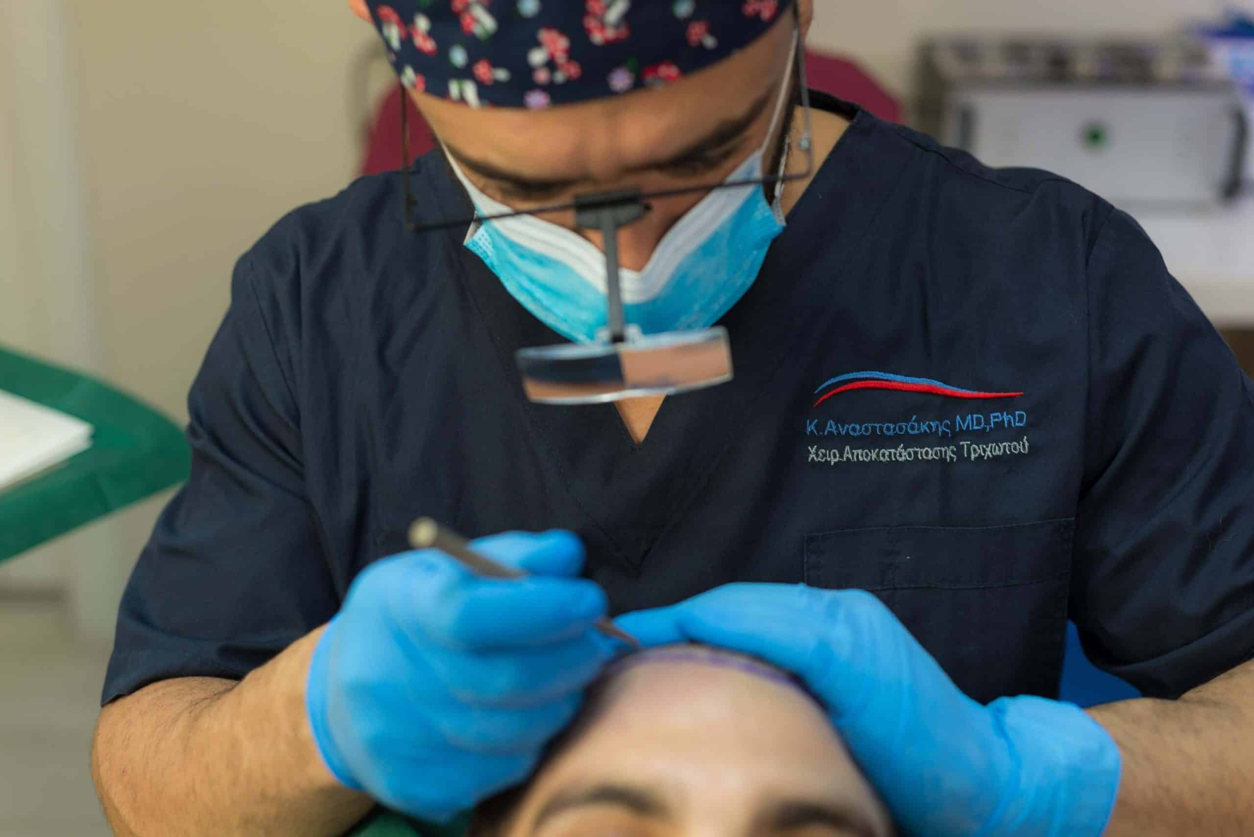
A key priority for the surgeon during hair transplantation is minimizing trauma to the blood vessels and nerves in the recipient area. Dr. Anastassakis explains how follicular unit transplantation can be performed with minimal trauma to the recipient site by using the appropriate techniques, ensuring an excellent aesthetic outcome for the patient.
Key Concepts
-
Trauma in the recipient area is exclusively iatrogenic. It is caused during the creation of thousands of recipient sites, and any damage to the scalp—particularly to the microvascular network—must be kept to a minimum so as not to compromise the survival and growth of the follicular units (FUs).
-
Each recipient site is three-dimensional, with width, depth, angle, orientation, and direction. The smaller the overall volume of the site, the less trauma to the blood vessels and scarring, and the more favorable the conditions for graft survival.
-
Instruments used to create recipient sites include fine-gauge hypodermic needles and custom-made disposable microblades measuring 0.5–0.8 mm in width, extremely thin, chisel-shaped, and with adjustable length.
-
The advantages of microblades are significant compared to needles in terms of graft survival, density, and naturalness of the result. Today, there are very few indications left for the use of needles in modern follicular unit transplantation.
Minimizing Trauma in the Recipient Area

The top priority for every surgeon in any procedure is minimizing surgical trauma, and hair transplantation is no exception.
The principle of “tissue respect” achieved through the minimally invasive techniques of the 21st century (FUT, FUE) could never have been accomplished with the mutilating procedures of “prehistoric technology” (punch grafts, scalp reductions, lifts, etc.).
The negative effects of extensive trauma to the recipient area were evident on many levels with the unreasonable procedures of the not-so-distant past.
Large wounds in the recipient area, such as those caused by 4 mm punches, damaged the vessels and sensory nerves of both donor and recipient sites, altering vascularization and scalp sensitivity. Meanwhile, scalp reduction, extension, and lift surgeries often left permanent damage across multiple scalp regions.
Fortunately, with FUT and FUE hair transplantation, trauma to scalp structures has now been minimized to its lowest possible level.
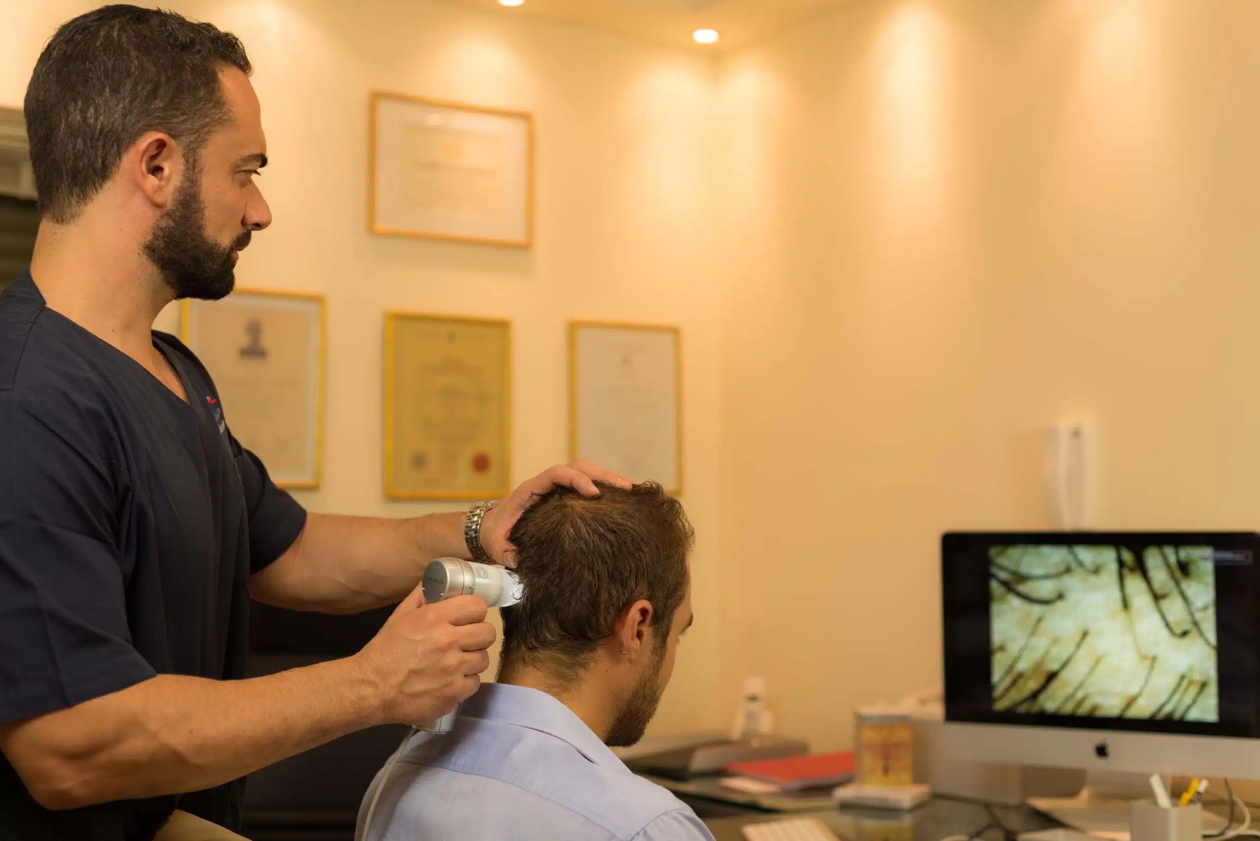
Scalp Vascularization
Under normal conditions, the scalp is exceptionally well supplied by the arterial plexuses of the skin. Its vascularization comes from branches of both the external and internal carotid arteries and from five pairs of arteries that traverse the subcutaneous fat.
The temporal and occipital regions are supplied by large branches of the external carotid artery, specifically the superficial temporal, posterior auricular, and occipital arteries. The frontal region is supplied by the supraorbital and supratrochlear arteries, which are small branches of the internal carotid artery, while the parietal region, which overlies the relatively avascular fronto-occipital aponeurosis, has a comparatively poor blood supply.
There are no arteries extending from the skull to the scalp, nor perforating branches from the underlying muscles to the scalp. It has been found that scalp vascularization is up to 10 times richer than in other anatomical regions of the skin surface of the human body. Anastomoses and collateral circulation in the scalp are so extensive, both ipsilaterally and contralaterally, that it has been reported that just two of the ten arteries (five pairs) are sufficient to supply the entire scalp. As a result, true anoxia and necrosis of the scalp are very rare complications. This fact provides a wide margin for surgical manipulation of the scalp.
However, the safety that this margin appears to provide is deceptive, since any vascular injury causes micro-disruptions in the perfusion of follicular units. Although the microcirculation of the scalp is extensive, it is inevitably disturbed during any skin surgery and therefore vascular trauma must be kept to the absolute minimum.
Because hair follicles are extremely sensitive to disruptions in blood supply and nutrition, protecting the microvascular network of the recipient area is of utmost importance. While the scalp itself will not undergo necrosis, the transplanted follicles in the recipient area may fail to survive if the microvascularization is significantly compromised.
The primary objective, therefore, is to keep trauma in the recipient area to the lowest possible level and limited strictly to what is absolutely necessary
How Is the Recipient Area Injured During Follicular Unit Implantation?

Injuries to the recipient area are exclusively iatrogenic and occur during the creation of the sites that will host the follicular units (FUs). Minimizing trauma in the recipient area first requires an understanding that each recipient site is three-dimensional, with the following characteristics:
-
Length
-
Width
-
Shape
-
Depth
-
Direction
-
Orientation
-
Angle
-
Placement
Because of this three-dimensional nature, each site has volume, even though on the surface it may appear as just a simple slit. It is also important to remember that we are referring to thousands of three-dimensional sites, placed in close proximity—often less than 2 mm apart.
The smaller the volume of each site, the less microvascular trauma per site, and the more favorable the “environment” for the graft to develop. Consequently, this significantly increases the chances of graft survival.
Key Considerations in Creating Recipient Sites
-
Size / Width of the Site: The pursuit of increasingly smaller recipient sites must take into account the “cost” of additional handling of the graft in order to fit it into the site.
-
Depth of the Site: Follicular units typically measure 3–5 mm in length. Recipient sites should not exceed 5 mm in depth, as greater depth risks damaging the subcutaneous vascular network of the scalp.
-
Density: Insufficient density will fail to provide aesthetic coverage, while excessive site density will irreversibly damage the subcutaneous vascular plexus of the scalp and compromise graft growth. Very high densities ultimately reduce graft survival and result in a thinner final appearance compared to initially lower densities.
-
Angle of the Site: The natural depth of scalp follicles is 4–5 mm, and their axis is always oblique to the skin surface, meaning the hair always emerges at an angle. The angle in each region of the recipient area is different and should replicate the angle of any remaining hairs, even vellus hairs in fully alopecic zones. It is important to remember that hairs eventually grow out at a slightly more obtuse angle than the implantation angle due to contractile healing. Sharper angles (≈15°) provide better coverage, while more vertical angles (≈75°) favor graft survival.
-
Direction: Defined as the orientation of the hair shaft as it emerges from the scalp. In the midscalp, it is generally posterior-to-anterior and should follow the orientation of surrounding hairs, including vellus hairs.
-
Flow: Refers to how the patient intends to style their hair, combined with the goal of achieving maximum coverage of posterior alopecic areas.
-
Orientation: Depending on the desired density and location of the recipient area, sites may be created in a sagittal or coronal orientation.
In addition to all the above, one must also consider the presence of existing follicular units in the recipient area, which must be protected both from direct trauma caused by the site-creation instrument and from damage to the subcutaneous microvascular network.
As early as 2001, Brandy published a technique for creating recipient sites in areas with pre-existing follicular units. His method required the use of 2.5–3.5× magnification and powerful Xenon lighting.
In an experiment conducted on his own patients, he divided the recipient area into two sides: on one side, the FUs were placed without magnification (naked eye), and on the other side, they were placed under magnification and enhanced lighting. The results became evident within the first 50 patients, and the experiment was discontinued for ethical reasons, since the magnified side consistently demonstrated significantly higher follicular survival and a notably lower incidence of telogen effluvium.
Recipient Site Creation Instruments
Recipient sites can be created with a variety of instruments. The “ideal instrument” is one that creates a site which:
-
causes the least possible tissue damage
-
has the appropriate depth to accommodate the follicular unit (FU)
-
has a shape that mimics the natural shape of the FU
-
has a shape that comfortably fits the FU
-
can be placed close enough to other sites to ultimately achieve the desired density
In modern hair transplantation (FUT, FUE), the use of advanced instruments allows us to achieve the same densities as in the past but with significantly less overall trauma. Alternatively, more sites can now be created—offering higher density—while causing a level of trauma to the recipient area comparable to that of older techniques.
Micro-blades
Most experts have now adopted the use of disposable, custom-made microblades, with varying widths (0.5–0.8 mm) depending on the size of the graft. These microblades are prepared prior to surgery using a special device that cuts Persona blades into the desired dimensions.
Microblades have a chisel-shaped edge, are extremely sharp, and allow for adjustable and highly precise control of the depth at which they enter the scalp. They also match the shape and length of the follicular unit (FU), enabling large hair transplant sessions (megasessions) with minimal trauma and mild postoperative edema.
The use of microblades creates microslit-type recipient sites and has the advantage of not altering the skin’s connective tissue, thus preserving the scalp’s natural elasticity. Each blade is typically used to create 100–200 recipient sites before being replaced
When a graft is inserted into a microslit recipient site, the remaining elasticity of the skin essentially “hugs” the graft (snug fit) and holds it in place, allowing grafts to be placed at higher densities.
Another advantage of using microblades is that once the graft is positioned in the site, it maintains full contact with the walls of the slit, enabling rapid restoration of blood supply and oxygenation.
Because there is no “dead space” between the graft and the site wall, scar tissue formation is minimal and healing is accelerated. Furthermore, since oxygen reaches the follicular unit (FU) through simple diffusion, the graft is evenly oxygenated and hypoxia is prevented. And because the FU is a compact anatomical entity, the occurrence of compressive phenomena after transplantation is also excluded.
Hypodermic Needles
The use of needles has remained as a “legacy” from older hair transplantation procedures with micrografts, which always included a portion of skin. This made it necessary to use some form of needle or other instrument that removed tissue in order to accommodate the graft in the recipient site.
However, due to their low cost and ease of use, needles continue to be employed by some hair transplant surgeons. Each needle is typically used to create 50–100 recipient sites before being replaced.
In contrast to microblades, the use of a needle to create recipient sites essentially acts as a micropunch, removing a thin cylinder of skin and thereby locally destroying elasticity. Even this microscopic excision of tissue significantly disrupts vascular continuity and blood supply, damages collagen, weakens the skin’s elastic support, increases scarring, reduces oxygenation, and delays healing.
By comparison, a simple incision made with a microscopic chisel blade causes far less damage than the excision produced by circular or elliptical micropunches or by 18G/19G needles. Even greater damage is caused by the use of lasers, which—fortunately—are no longer employed in modern hair transplantation.
Problems Caused by the Use of Inappropriate Instruments
The use of inappropriate instruments can result in graft “popping”, where grafts are more easily pushed out of the needle-created recipient sites. This occurs because the upper portion of the epidermis—which would normally help hold the graft in place against the elastic “pressure” of the recipient area—is absent
The forced repositioning of a graft is often traumatic and subjects the graft to additional handling, from which it may not always survive to grow successfully. With the use of needles, a portion of skin is removed, and the contact between the graft and the recipient site walls is imperfect. As a result, oxygenation is suboptimal, and the formation of scar tissue between the graft and the site walls is more extensive.
The result is that the recipient area cannot “hold” the same number of grafts in a single session, since the grafts tend to be expelled from the opening (popping) or to “float” within it. Postoperative growth also tends to be inferior, as the additional scar tissue is avascular, reducing overall blood supply per unit volume of skin. Consequently, the aesthetic outcome is often below expectations. Finally, the formation of postoperative micro-crusts is more pronounced with the use of needles, and healing is slower.
One detail worth noting, however, is that if the needle is inserted only up to the bevel and not to the full round section, the recipient site ultimately has a crescent shape, and the needle does not act as a micropunch. Still, it is not easy to maintain such precision and consistency across thousands of sites.
Despite their significant disadvantages, it should be acknowledged that there are certain indications for the use of needles in recipient site creation, such as very large follicular units or curly hair. The group of Yamamoto et al. reported a 10-month survival rate of 102.2% with micropunches, compared to 98.9% with microslits in patients with curly hair. Innovative solutions have also been proposed for the continued use of needles in hair transplantation—for example, the use of epidural anesthesia needles, which, due to their angled tip, create recipient sites that reduce popping.
Nevertheless, the author strongly recommends the use of microblades.
SUMMARY:
Respect for tissue must always be the surgeon’s top priority, and minimizing trauma to the vessels and nerves in the recipient area cannot be overstated.
While each recipient site is microscopic, when one adds up the cumulative iatrogenic damage from thousands of sites, it becomes clear that small differences in technique or instrumentation translate into significant differences in overall trauma.
Today, the use of disposable custom-made microblades is the recommended technique for creating recipient sites, as they cause less damage, ensure higher graft survival, and deliver superior aesthetic outcomes compared to the older use of needles