

Donor Area and Safe Donor Zone (SDZ): Key Concepts
The safe donor zone is defined as the anatomical area that contains only hormone-independent follicular units, and only these may be harvested from the donor area. There are clear landmarks and guidelines for assessing the safe donor zone, and measurements must be precise, with any overextension strictly avoided.
The donor area as a whole possesses unique characteristics in every patient, which must be considered in relation to the requirements of the recipient area for grafts. In general, up to 50% of the donor area may be transplanted in total. The donor area could be described as the most important region of the scalp in patients with hair loss—excluding the anterior area, which is by definition devoid of hair in those seeking transplantation.
The characteristics of the donor area are what make hair transplantation procedures feasible, and they must be thoroughly evaluated before a patient is deemed suitable and proceeds with follicular unit transplantation. The principal characteristics include:
-
-
The width of the safe donor zone
-
Laxity
-
Glidability (mobility of the scalp over the underlying structures)
-
Elasticity (or extensibility)
-
Presence of previous scars or trauma
-
Follicular density and calculated density
-
Hair type (straight, curly, etc.)
-
Hair color
-
Hair shaft diameter
-
All these elements must be integrated, and in conjunction with the number of grafts required to achieve satisfactory and natural coverage of the bald areas, the precise surgical plan to be followed must be determined. Failure of the surgeon to recognize the particular characteristics of each patient’s donor area will not necessarily result in complete failure of the hair transplantation, but it will certainly diminish its effectiveness and, consequently, the patient’s satisfaction.
In the sections addressing the donor area, the following topics will be extensively analyzed:
-
The safe width of the donor zone, ensuring that all follicular units harvested are indeed hormone-independent
-
The characteristics of the scalp in the donor area
-
The details of donor density and its remarkably interesting geometric and mathematical aspects
-
Closure techniques designed to achieve minimal tension and scarring in the donor area
-
Potential complications in the donor area, their causes, and the key guidelines necessary for their prevention
Important Note
For a successful hair transplantation, the follicular units transplanted must be hormone-independent, so that they do not shed in the recipient area under the influence of androgens. The so-called safe donor zone is defined as the anatomical area containing only hormone-independent follicular units.
It is considered an axiomatic principle that the follicular units of the safe donor zone are hormone-independent, meaning they are not miniaturized by the action of androgens, and this property is preserved even after their surgical transfer to the hormone-sensitive recipient area.
However, the number and distribution of follicular units within the safe donor zone (as in any other area) are genetically determined and remain stable throughout life. The safe donor zone is, therefore, the “capital–heritage” of the patient with hair loss, and the hair transplantation surgeon is essentially entrusted with “investing” it in the best possible way, since no method has yet been found to increase it.
The Width of the Safe Donor Zone
When the hair transplantation surgeon decides to operate on a patient with hair loss, there are three factors of utmost importance that must be ensured:
-
The follicular units to be harvested must contain permanent, hormone-independent follicles.
-
Any deficit created—if the FUT technique is employed—must be closed without tension at the wound edges and with an inconspicuous scar.
-
The removal of the graft must cause minimal collateral loss of adjacent follicular units.
The width, length, and surface area of the donor region are not identical for every patient; however, the anatomical boundaries of the safe donor zone tend to be similar across patients and are defined by clear anatomical landmarks. The total surface area of the human scalp is approximately 520 cm², and the area of the safe donor zone that may be harvested has been calculated to represent about 12–15% of the total. Each donor area possesses specific and unique characteristics that provide the surgeon with essential information in order to adjust the patient’s expectations to the true capacity of their donor area.
Alt was the first to define the exact boundaries of the safe donor zone. Specifically, an imaginary vertical line drawn in front of the external auditory canal delineates the anterior boundary of the safe donor zone. At this point, its width measures 6.5–7 cm, and as it extends toward the occipital region, this width gradually decreases.
The superior boundary of the safe donor zone in the occipital region is located where the horizontal extension of an imaginary line, beginning 1–2 cm above the ear, intersects with the mid-occipital line. A margin of at least 2.5 cm is recommended above the upper limit of the donor area, in order to ensure coverage of scars from future procedures.
The inferior boundary of the safe donor zone is not fixed and is determined through the physical examination of the patient and the family history, since follicular loss with aging may also occur in the scalp region adjacent to the nape of the neck.
defined as any area that retains more than 8 hairs within a circular area of 8 mm² in individuals with alopecia at stages III–VII. As expected, this zone is significantly “narrower” than that described by Alt, although it begins 2.5–2.8 cm more anteriorly.
An interesting finding from Unger’s study on 328 men over the age of 65 was that more than 78% of men never reach stage VII, indicating that the onset of hair loss at a young age does not necessarily progress to advanced-stage alopecia, but only in about 20% of these men.
Finally, Avram suggested that the region bounded inferiorly by the occipital protuberance and superiorly by an imaginary line connecting the auricular helices circumferentially is the safest zone for graft harvesting.
Donor Area Evaluation Techniques
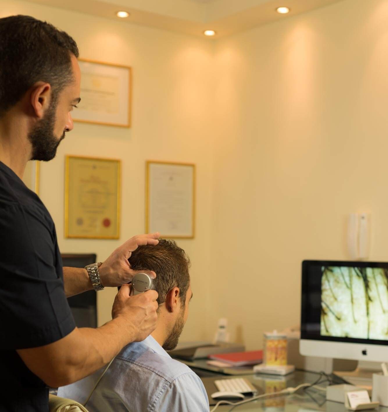
A thorough clinical examination of the scalp is essential before every hair transplant procedure, in order to identify possible signs of future progression of hair loss to other regions. The examination is typically performed with the patient’s hair wet, as this allows the surgeon to more easily detect the classic features of alopecia—most notably the visibility of the scalp skin between hairs, which results from follicular unit miniaturization.
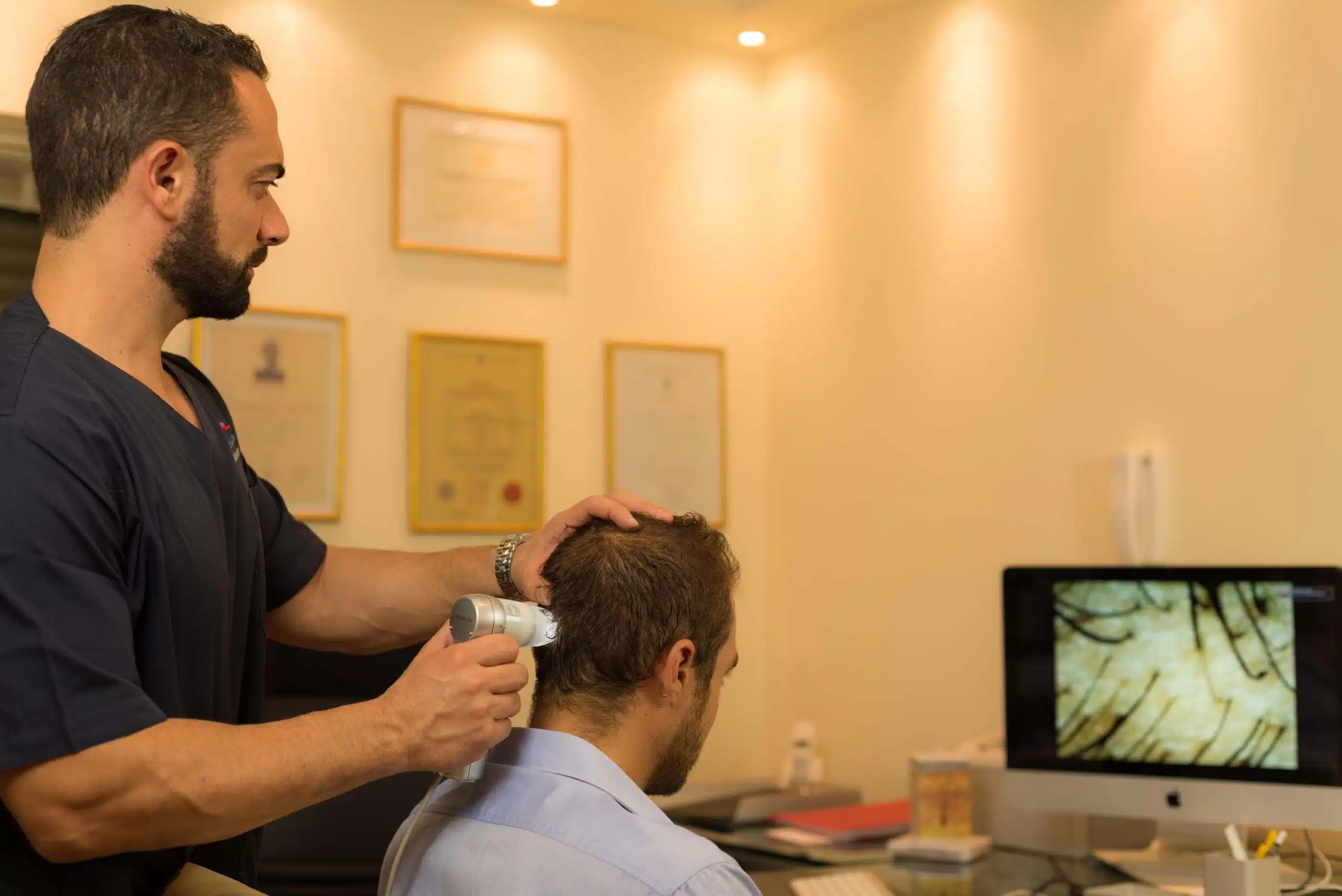
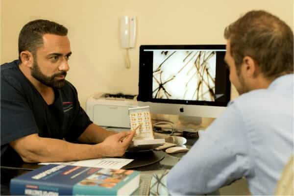
The next crucial step in donor area evaluation is the measurement of miniaturization in both the donor and recipient zones. Assessment of follicular miniaturization in the occipital donor area is carried out using a magnifying video dermatoscope (ideally Proscope HR2). A high degree of miniaturization in the superior occipital zone may indicate a potential reduction of the safe donor area as hair loss progresses. Conversely, significant miniaturization across the wider donor region almost always suggests a progression towards Norwood stage VII baldness.
It is important to note that the younger the patient, the more challenging it is to predict the future course of hair loss. On the other hand, the older the patient, the more reliable the surgeon’s assessment becomes regarding the long-term stability of the donor area and the overall success of the hair restoration surgery.
When determining the width of the strip graft that can be safely removed from the donor area during a hair transplant, the main limiting factor is the avoidance of creating an overly sparse appearance in the donor region. In other words, the surgeon must remove such an area of scalp tissue that the remaining hair density does not give the impression of thinning.
It is therefore generally accepted that if up to 50% of the donor area is removed over multiple sessions, this risk does not exist. If the hair characteristics are favorable, this percentage can be increased even further. Conversely, if the patient’s hair is fine and the density low, the percentage must be reduced significantly, and in some cases, the individual may not even qualify as a suitable candidate for hair restoration surgery.
Furthermore, the length of the strip graft that can be excised is not fixed. The pericranial distance between the ears varies between 25–32 cm among individuals. Even in the same patient, the exact length and width of the excision is determined by the requirements of each session and the overall treatment plan.
For relatively small sessions (<1,200 FUs), many surgeons prefer asymmetrical incisions, harvesting from only one side of the safe donor zone, thereby leaving the opposite side “untouched” for a future session. For medium sessions of 1,500–2,500 FUs, most surgeons opt for a full “ear-to-ear” incision, typically less than 1 cm in width. In the mastoid regions, however, the strip must be narrower due to reduced scalp elasticity.
Further details on graft extraction using the FUE (Follicular Unit Excision) technique are discussed extensively in the corresponding section.
Summary
he donor area is the true “inheritance” of every hair transplant patient. For this reason, the surgeon must handle it with the utmost respect, ensuring prudent use and avoiding unnecessary depletion of its resources—or his own technical skills. Every follicular unit (FU) is valuable, and every effort must be made to minimize trauma to the donor area, the follicles, and the vascular and neural networks of the scalp.
Scalp Laxity = Elasticity + Mobility
The terms laxity, elasticity, and mobility of the scalp are often incorrectly used interchangeably, but they describe different properties. According to Seery, laxity depends on two distinct factors:
-
Mobility: The ability of the scalp to “glide” over the pericranium. This property is unrelated to skin elastin or the ability of the scalp to stretch and is more likely determined by the density of fibrous tissue beneath the galea aponeurotica. It can be compared to sliding a carpet across a wooden floor, where the objects on the carpet do not move, nor does the carpet expand, but it shifts as a whole.
-
Elasticity: The ability of the skin to stretch, mainly dependent on the elastin content of the dermis. A useful analogy is the stretching of a plastic food wrap, which increases in surface area as tension is applied.
The higher the values of both properties, the wider the strip graft that can be harvested from the safe donor zone without creating excessive tension or tissue distortion. Mobility was historically more important in reduction surgeries, scalp lifts, and scalp extensions, whereas elasticity is a key parameter in modern strip excision hair transplant techniques.
Measuring Donor Area Laxity
Measuring the overall scalp laxity is crucial when a strip graft wider than 10 mm is planned. Norwood initially suggested compressing the scalp bilaterally from the temporal areas toward the midline and observing the resulting skin folds. The scalp was then classified as tight, moderately tight, slightly tight, average, slightly loose, moderately loose, or loose. While widely used for years, this method is subjective and unreliable.
Other methods were later proposed—by Bosley (1980), Feldman, and more recently Mohebi, who invented the Laxometer. In addition, Mayer & Pauls introduced an objective, reproducible technique, demonstrating that wound tension of up to 5 Newtons (500 g) is associated with scars only 1–3 mm wide, highlighting the importance of keeping postoperative wound tension below this threshold.
For optimal healing, incisions in the donor area must follow the natural Langer’s lines of minimal skin tension, first described in 1881 and later updated by Cox in 1941. These lines are believed to result from the arrangement of collagen fibers in the dermis. On the scalp, they run vertically across the superior regions, gradually becoming horizontal in the occipital area.
Not all anatomical regions of the scalp have equal laxity. The retroauricular area shows the lowest laxity, followed by the mid-occipital zone, while the occipital region is the loosest and therefore the most “profitable” donor site. The ideal shape of the elliptical excision in strip FUT surgery reflects these variations: narrower above the auricular helix, wider in the occipital zone, and narrower again as it approaches the opposite ear.
Written by Dr. Konstantinos Anastassakis
 0
0