

Graft survival is a key factor in the success of hair transplantation. Read everything you need to know..
Graft Survival: Basic Concepts
- In modern hair transplantation, sessions last longer, grafts remain outside the body for several hours, and they are exposed to various sources of mechanical and biochemical stress (dehydration, mechanical trauma, etc.).
- Fortunately, many techniques, interventions, and technical tools have been developed to counter almost all of these stress factors.
- High-level expertise, attention to detail, focus, care, and dedication from the surgeon and the team are essential to achieve high graft survival rates (>95–98%).
The second stage in hair transplantation is the placement of grafts into the recipient area, which is divided into the creation of recipient sites and the actual insertion of grafts into those sites. However, before reaching this stage, the surgeon must ensure optimal conditions for graft survival both before and during implantation.
Graft Survival: A Matter of Life and Death!
As already mentioned, in both FUT and FUE techniques, strictly follicular units (FUs) are transplanted, exactly as they are obtained—either from the strip graft (FUT) or through direct extraction from the scalp (FUE). These FUs contain a varying number of follicles, typically 1–4, while the occurrence of FUs with 5 or 6 follicles is extremely rare.
The evolution of hair transplantation and the establishment of the FUT technique significantly reduced the size of grafts while dramatically increasing the number of grafts that can be transplanted in a single session. This also raised the achievable density in the recipient area to levels unimaginable just a decade ago.
At the same time, procedure durations have lengthened, and during both the intraoperative and postoperative periods, each graft is exposed to multiple sources of mechanical and biochemical stress—any one of which alone would be enough to significantly reduce its survival chances.
Factors Affecting Graft Harvesting
To achieve high density and coverage with the FUT hair transplantation technique, it is essential first and foremost to ensure maximum yield from the donor area.
The mistakes of the past in all aspects of hair transplantation were dramatic, and gaining knowledge through experience was a slow and painful process. The following conditions are necessary to maximize graft yield:
-
Use of a single-blade knife
-
Use of magnification and continuous visual control during donor strip excision
-
Highly trained staff for graft preparation
-
Use of a microscope during graft preparation
-
Preparation of grafts on an illuminated surface
-
Preservation of grafts in the form of follicular units (FUs)
-
Preparation of FUs with sufficient surrounding tissue to provide protective support to the follicle (“chubby” grafts)
-
Maintenance/storage of grafts until placement under conditions that minimize losses from anoxia or dehydration
If any of these conditions are not met, one cannot rely on the technical advantages of FUT to achieve good results. It is futile to apply perfect placement techniques in the recipient area when 30% of grafts have already been destroyed due to the use of multi-blade knives or poor handling during preparation.
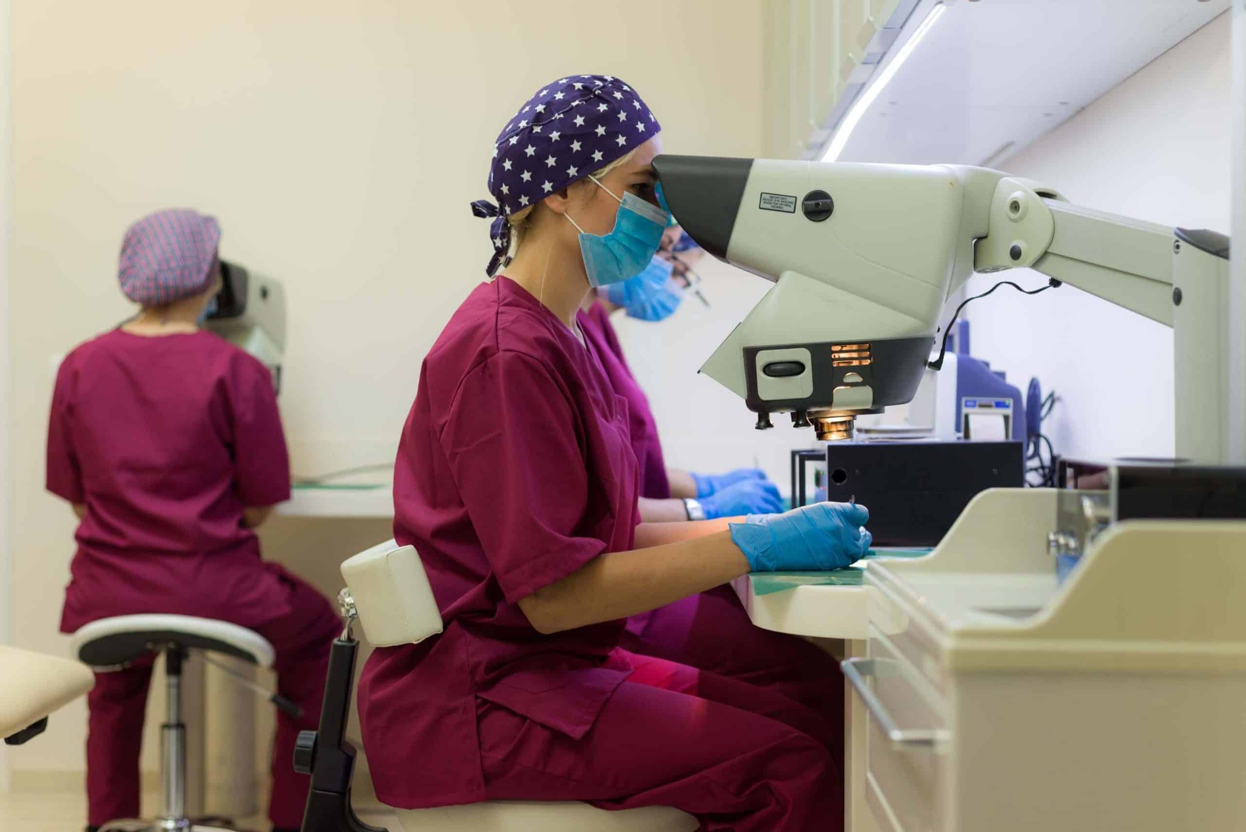
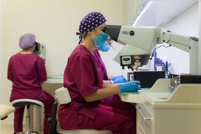
Nevertheless, not all surgeons have adopted the use of microscopes in their practice, and some objections to the obvious logic of microscope use border on “phobia”—especially when extensive and significant studies have demonstrated superior survival of FUs processed under magnification.
Factors Affecting Graft Survival
Let us first examine what can cause reduced graft growth from the moment they are prepared:
- Dehydration: From the moment the donor strip is removed until each FU is placed into the recipient site, grafts remain exposed under intense surgical lights and in an air-conditioned/dry/warm environment. Smaller FUT grafts have a higher surface-to-volume ratio, making them more prone to dehydration. Their small size also makes insertion more difficult, meaning they often remain in the technician’s hand longer before being placed. Studies have shown that even 3 minutes outside the Petri dish is enough to significantly reduce FU survival.
A study by Gandelman et al. showed that dehydration has the most severe negative impact on FU survival compared to any other factor.
- Mechanical trauma: Inserting thousands of tiny FUs into equally tiny recipient sites is technically demanding, and the risk of injury is high. When using the traditional forceps method, extreme care is needed. Jewelers’ forceps are commonly used, but the pressure exerted by the fine tip can literally crush graft tissue. The pressure per unit area can be compared to the heel of a stiletto shoe exerting more pressure/cm² than an elephant’s foot. The less protective tissue surrounding the follicle, the higher the risk of irreparable damage. Single-hair FUs are 40% more vulnerable than multi-hair FUs. Trauma may also occur from repeated attempts to insert grafts into sites, especially by inexperienced technicians. The most critical damage occurs when the bulb or bulge region is compressed. Shapiro estimated that 90% of poor graft survival cases are due to mishandling trauma.
- Out-of-body time: With the rise of megasessions, even experienced teams sometimes keep grafts outside the body for >9 hours. Limmer showed in 1996 that, under ideal conditions, after 6 hours out of the body, graft survival decreases by 1% for each additional hour.
- Ischemia-reperfusion injury: During FUT and FUE, follicles are separated from their blood supply, causing temporary ischemia/hypoxia. ATP breakdown halts at the hypoxanthine/xanthine step due to lack of oxygen, leading to intracellular accumulation. When reperfusion occurs, xanthine oxidase rapidly converts these into uric acid and releases a surge of free radicals/ROS, overwhelming antioxidant defenses and leading to apoptosis and cell death. Although transplanted follicles oxygenate passively via diffusion for the first 3 days and revascularize on day 4, Cooley showed that they still display a 200–600% increase in malondialdehyde (MDA), an indicator of reperfusion injury, possibly linked to reduced survival.
- Popping: Occurs when the pressure exerted by surrounding sites pushes previously placed grafts out. This typically happens when grafts are too large for the sites or when sites are placed too densely. Re-insertion causes unnecessary mechanical stress, reducing survival. Excessively deep sites can also cause bleeding that pushes grafts upward.
- Excessive depth: “Sinking” of grafts below the epithelial surface (due to insertion or pressure for hemostasis) can result in lower final density, epithelial cysts, or infection.
- Too shallow depth: Grafts placed too superficially can cause a “tenting” effect, where the scalp epithelium rises to cover the follicle’s dermal portion. Rarely, this can cause graft loss.
Bending & Piggybacking:
-
Bending occurs when grafts are pushed into sites rather than gently pulled, causing the dermal papilla to face upward. Some believe this may cause “curly” hairs, but outcomes are uncertain.
-
Piggybacking occurs when a graft is mistakenly placed on top of another in the same site, leading to cysts and ingrown hairs.
Methods developed to minimize these risks:
-
Dehydration: Use cold LED surgical lights, maintain low temperature and high humidity in the operating room, keep grafts continuously in Petri dishes until implantation, frequently moisten grafts with saline when outside, minimize time outside solution, and use chilled storage medium.
-
Mechanical trauma: Ensure safe graft size with adequate perifollicular tissue (medium or chubby grafts), match site size/depth to graft size, handle from non-vital areas, minimize re-insertion attempts, and maintain high staff training standards.
-
Out-of-body time: Use specialized storage solutions, add Vitamin B12 and aminoguanidine to storage medium, particularly in megasessions.
-
Popping: Ensure proper graft sizing, match site to graft to avoid compression or “floating,” maintain adequate spacing, and ensure correct site depth.
-
Ischemia-reperfusion injury: Use antioxidant-containing storage solutions, speed up graft preparation/placement for faster revascularization, avoid strong antiseptic solutions.
-
Bending & Piggybacking: Continuous monitoring of intraoperative bleeding, improved lighting and magnification, use of dyes to mark recipient sites, and use of implanters.
What else can cause reduced graft survival?
All the factors associated with poor or below-expected follicular growth after transplantation that are related to the above-mentioned conditions are referred to as H-factors (Human factors).
Greco divided them into two categories:
-
Primary H-factors: factors related to graft preparation at any stage of hair transplantation—from the donor incision and strip preparation to graft popping due to incorrect placement.
-
Secondary H-factors: all predisposing factors that can indirectly injure grafts. These include a dull blade, poor lighting, inadequate hemostasis, FU dehydration, antiseptic action, mismatch between site size and graft size, and excessively dense graft placement.
Factors linked to poor or below-expected follicular growth that are related to unknown causes are referred to as X-factors.
The term X-factor was first introduced by Norwood and Shiell to describe a set of variables that caused unexpected and unexplained poor growth in 4 mm punch grafts—factors that varied unpredictably among patients. Later, they suggested that a rejection mechanism might be responsible, leading to local ischemia and eventual graft atrophy. However, this theory was not sufficiently proven. Cooley and Vogel later demonstrated that the X-factor was essentially the result of transection of the dermal papilla during graft preparation or placement.
Thus, if we adopt the view of Cooley and Vogel, both X-factors and H-factors ultimately depend entirely on the quality of the surgical team’s work.
Even very small differences among teams—in training, attention, and dedication—can result in major differences in final outcomes. The field of hair transplantation is undergoing constant micro-evolution, which creates continuous pressure for ongoing training of surgeons and assistants.
Such continuous training is essential, as it increases productivity and confidence, improves emotional stability and concentration during repetitive micro-tasks such as graft preparation, and strengthens teamwork and cooperation under stressful conditions.
Only under these conditions can high-level densities and superior graft survival be achieved in hair transplantation procedures.
Graft Survival: Summary
Techniques and innovations aimed at reducing the biochemical and mechanical stress that grafts undergo during preparation are now applied successfully, and achieving survival rates of >95–98% is the target in routine hair transplantation.
With regard to graft survival, when the surgeon performs the donor incision or FU extraction (in the FUE technique) correctly, when graft preparation is carried out carefully by experienced technicians, and when graft placement follows simple and clear principles, the outcome of hair transplantation is almost always guaranteed.
It is the surgical team as a whole that ensures the excellent results of modern hair transplantation, and its members must always work with the highest levels of attention, focus, and dedication.
Written by Dr. Konstantinos Anastassakis
 0
0